


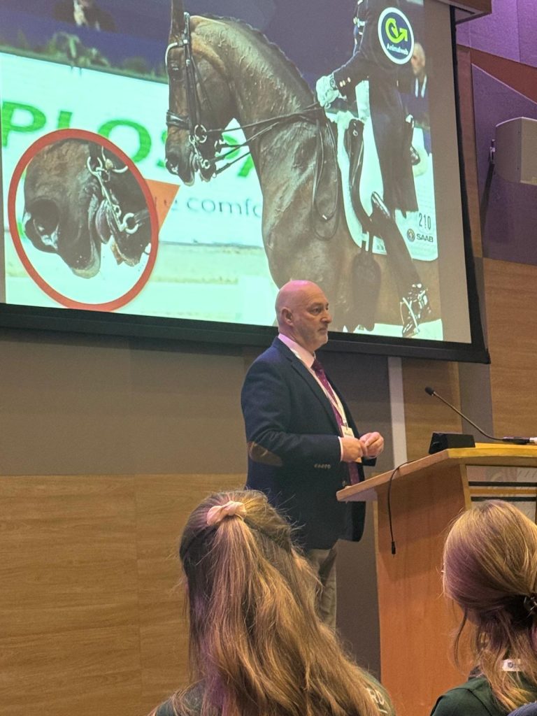
At this morning’s British Equine Veterinary Association (BEVA) Congress in Birmingham, Dr David Marlin presented fascinating new research exploring the phenomenon of the so-called “blue tongue” in horses.
Images of horses with discoloured tongues have been widely circulated on social media in recent years, sparking debate about what causes this change and what it might mean for equine welfare. Key questions include: How common is it? Does it cause pain? Is the bit to blame? Could oral conformation play a role?
To investigate, Dr David Marlin and his team developed a standardised photographic method to measure tongue and gum colour in horses before and after exercise. Using consistent lighting, camera settings, and a colour reference card, they were able to analyse images using the red–green–blue (RGB) colour system and calculate a red–blue ratio, which indicates relative oxygen saturation in the tissue.
The study followed nine non-elite horses ridden in walk, trot and canter for 30 minutes in snaffle bridles with varying noseband tightness (two fingers, 1.5 fingers, and one finger). Photographs were taken before exercise and within one minute afterwards, on both sides of the mouth, with a veterinary surgeon ensuring no oral lesions were present.
Findings:
- No significant differences were observed between the left and right sides of the mouth.
- No significant differences between upper and lower gums.
- The tongue was consistently lower in red and blue content compared to the gums, resulting in a higher red-blue ratio.
- Importantly, no significant changes in tongue or gum colour were detected after low to moderate intensity exercise under the conditions tested.
Dr Marlin concluded that while this pilot study suggests short periods of ridden exercise in a snaffle with varying noseband tightness do not significantly alter tongue colour, further research is needed. Higher intensity exercise, longer durations, different bits, or tighter nosebands may produce different results. He also noted that while photographic colour analysis provides an objective method, it is not a direct measure of tissue oxygenation, and complementary non-contact techniques such as imaging photoplethysmography (iPPG) may offer further insight.
This research provides an important step towards understanding the “blue tongue” phenomenon and its potential implications for horse welfare.
There is NO CPD Certificate for this video.
Transcription
Here is a transcription of the webinar. (Please note this is an automated transcription, so we apologise for any errors.)
Hi there, I’m David Marlin and this is the presentation that I gave at the British Equine Veterinary Association Congress in Birmingham this morning. And it’s entitled, Does Ridden Exercise Influence Tongue Colour? A Standardised Photographic Approach. So many people be aware that images like this have started to appear more commonly ⁓ on various social media and in the press over the past few years
And this is what’s become known as blue tongue. But there’s a lot of questions we should be asking about blue tongue. What is it? What causes it? How common is it? And these are just some of the questions here. Does it cause pain? What role does the bit play? ⁓ What about the role of oral confirmation? ⁓ Do blue tongues occur in wild horses? So we’ve actually got lots of questions and probably some of the most important are what causes it and how common is it?
But when we’re trying to take images of the equine tongue, we need to consider there’s a lot of factors that can influence the color balance in digital photos. And I’m not going to go through all of these here, but you can read them there. Cameras have what are called white balance settings. Different cameras will produce different tones. The distance you are from the subject, what the background is, all of these factors and more can affect the actual
final colour of the image. So what we set out to do was to try and produce a standardised approach to imaging so that we could actually compare the colour of the mouth, including the tongue and the gums of horses, ⁓ looking at say difference between left and right side, looking at whether there was difference in the tongue and gums and also looking at the effect of exercise.
And to do this, this standard approach, the main thing here probably is that we use a standard distance, a standard lens, standard camera settings, but also this light ring, which is 5,600 Kelvin, which is considered to be daylight balanced light. And because we’re using that close to the horse, that essentially controls the color of the image.
Now, at the same time, we can also use a color reference card so that we can correct the colors of the images so that they’re all standardized after they’ve been taken. And the way we’re analyzing this is based on the RGB system, which is red, green and blue. So, for example, this is between zero and two five five for each of these colors. ⁓ White is two five five, two five five, two five five, whereas black is zero zero zero.
A ⁓ full red intense red is 255 red with no green and no blue whereas a full blue color would be a zero for red zero for green and 255 for blue so any color is made up of combinations of red green and blue on this scale of nought to 255 So if we look here, we can see that different colors of blood are made up by
a different combination of red, green and blue. So for example, 100 % oxygen saturated blood is around, as you can see there, 220, 30 and 20. And when we look at this on a graph, we can see that the as the oxygen saturation decreases, so does the amount of red decrease.
but as the oxygen saturation decreases, the amount of blue also ⁓ increases. And the tissue coloration is dependent on the color of the tissue itself, but also the blood flow or blood volume within that tissue and the oxygenation of the blood. Now, what we can do is the absolute amounts of red and blue may change with imaging factors and blood flow and volume.
but the ratio of red to blue is independent of these and represents the relative degree of saturation. As you can see here, the red-blue ratio is represented by the purple curve in this image. So what is the basis for using colour to assess ⁓ the oxygenation of tissues? Well, as tissue colour cannot change in the short term,
any change in colour will be related to changes in the blood volume or blood flow and or changes in the oxygenation of that blood, provided the imaging is standardised. So we can use this to try and compare, for example, here the tongue, the lower gingival mucosa and the upper gingival mucosa. So in this study, which was covered by ethics, we had nine non-elite horses and riders.
⁓ They were horses were ridden in walk trot and canter for approximately 30 minutes in a snaffle with a nose band originally starting at two fingers then at 1.5 fingers and then at one finger and we obtained images before exercise and within one minute of horses finishing exercise and also we imaged the left and the right side. horse’s mouths were exposed ⁓ for
photography by a veterinary surgeon and none of the horses in the study had oral lesions. In terms of the processing, you can see here on the right an example of an image with the processing applied. We used image J. The region of interest, in this case the tongue, was traced. We were careful not to include any pigmented areas. Analysis was done with the colour histogram plug-in for image J.
which is a standard plugin that’s available. Correction, then for color reference card, colors red and blue and white in Adobe Lightroom. And then we calculated the red pixel mean, the blue mean, and the red-blue ratio from those.
In terms of statistics, the data were not normally distributed, so we applied a box Cox transformation. We then followed this with analysis of variance with the linear mixed effects model with horse as a random effect and time side and location as fixed effects. If significance was reached less than 0.05, then we applied post hoc to these tests to compare means. So.
This just shows a set of data, a set of images from one horse. In terms of where there were significant differences, we saw that there was a significant effect of location in that the red content or the blue content or the red-blue ratio were all different between the, not different between the upper and lower
Gingiver as we would expect but they were they were similar but the tongue was lower in red and Was also lower in blue but relatively this gave a higher red blue ratio for the tongue but There were no significant effects of horse There was no difference between the left and the right side And there was no effect of time
So I time here means exercise. And this is just showing the red content as an example. So this was the most significant effects of red content, blue content or red blue ratio. So obtaining images with a standardized approach, correcting to a color standard and processing as described in this group of nine horses, indicated no differences due to the left or right side or the upper or lower gingival mucosa.
The tongue was significantly different from both the upper and the lower gingival mucosa. So in this small group of horses, a short period of low to moderate intensity exercise in a snapper bridle with noseband tensions from two to 1.5 to one finger did not produce any significant change in tissue color with this technique. So limitations for discussion. Recording color change is not a direct measurement.
tissue oxygenation, but it does reflect any change will reflect tissue oxygenation and or blood flow. It’s quite possible that higher intensity exercise, higher rain tensions and different bridles may produce different results with respect to pre and post exercise. We’ve looked at already at other methods of trying to get at the tongue and the tissue oxygenation, the mucosa, but pulse oximetry
contact pulse oximetry, whether transmission or reflectance is not an option for direct measurement in conscious horses. But it’s possible that other non-contact techniques such as IPPG could be suitable for measurement during written exercise. So what we have here is a standardized approach that would allow you to objectively compare the color of the mouth in terms of the tongue and the gums.
before and after exercise to see if there actually has been a change in the coloration using this standardized approach.
