PRESENTED BY Jennifer Stewart, BS, DVM, DACVP, MRCVS. Animal Health Trust, Newmarket, UK (In the Defra Report 2014)
Acute ragwort poisoning, though uncommon, may be seen histologically, ranging from multifocal to extensive necrosis without further injury. Haemorrhage associated with the hepatic necrosis is common. The distribution of hepatic necrosis in these cases resembles that caused by numerous other hepatotoxins, and is therefore non-specific. In acute toxicities caecal and colonic oedema and infarction may also be observed at post-mortem examination in some cases.
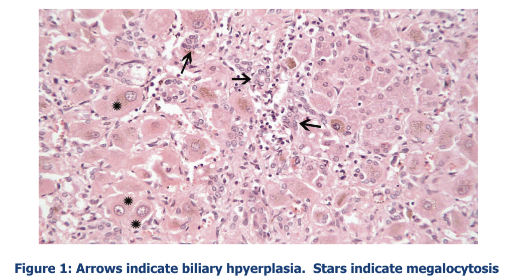
Chronic toxicity results in the more ‘classical’ presentation of bridging fibrosis, nodular re-generation, biliary hyperplasia, and megalocytosis. megalocytosis appears as enlarged hepatocytes and nuclei. This feature is a hallmark of pyrrolizidine alkaloids, whose toxic metabolites bind to DNA and prevent successful mitoses. Megalocytosis was found experimentally to develop over the course of 14 weeks following two weeks’ exposure. By six months post-exposure, the megalocytosis was considered the most prominent feature, and is the feature most associated with pyrrolizidine alkaloid toxicity. Regenerative nodules are typically small due to the lowered number of hepatocytes capable of successful mitosis. Similarly, biliary hyperplasia is anticipated to represent a response to the general regenerative stimuli.