


Rachel Gough MA MA VetMB CertAVP (EM&ESM) DipECEIM MRCVS – Specialist in equine internal medicine
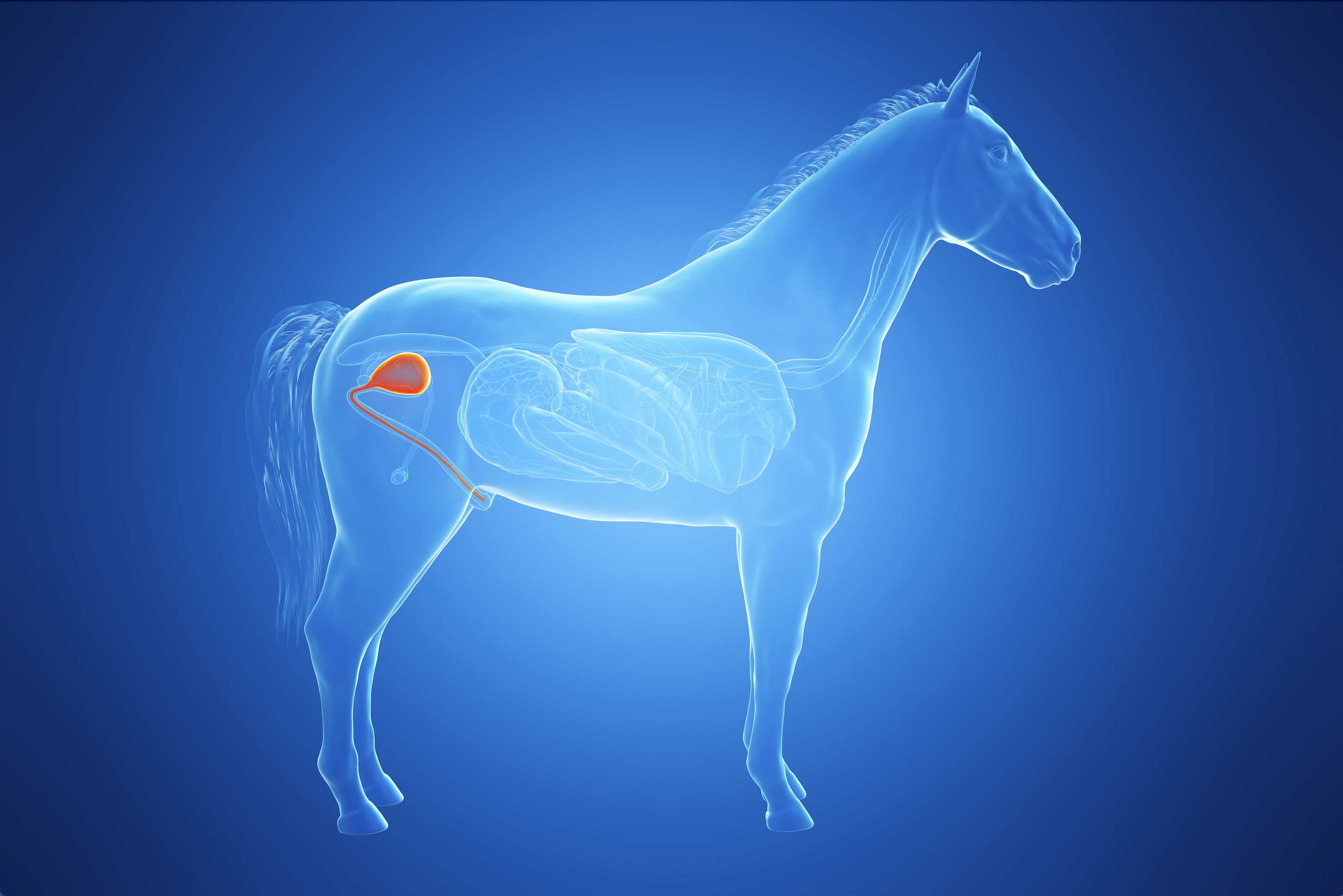
Compared to the gastrointestinal or musculoskeletal systems the urinary tract infrequently causes problems in adult horses. An uncommon but important issue that can arise is bladder stones. These are more correctly referred to as cystoliths or cystic calculi. “Cyst” derives from the Greek for bladder and “lith” from the Greek “lithos” meaning stone; calculi is from the Latin for pebble.
When should I worry that my horse might have a cystolith?
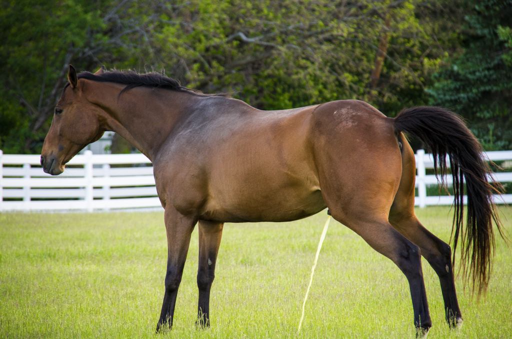
Cystoliths typically cause stranguria which means difficulty urinating. It manifests as a horse repeatedly posturing to urinate with little or no urine expelled; in males the penis is often repeatedly extruded and in females the clitoris everted. In the case of cystoliths, stranguria most commonly occurs in association with exercise, likely due to agitation of the stone within the bladder. It is important to note that standing stretched out as if posturing to urinate also occurs with generalised abdominal discomfort; it is not uncommon for horses with gastrointestinal forms of colic to perform this behaviour. Another classic sign of cystoliths is haematuria (blood in the urine), again most commonly in association with exercise. This can be microscopic and only detected on a urine sample or the urine may be visibly bloody. The urine will typically be red throughout the stream, not just at the beginning or end. Other signs include pollakiuria (the frequent passage of small volumes of urine), urinary incontinence (dribbling) or recurrent colic. None of these signs are specific for cystoliths so it is very important to discuss any concerns with your vet!
What are cystoliths made of?
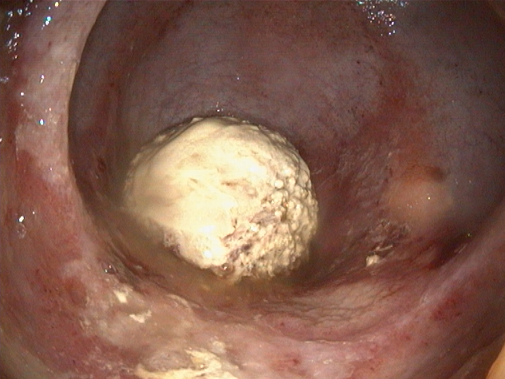
In horses, cystoliths are primarily composed of calcium carbonate. They are classified as type I and II. Type I are usually ovoid in shape, yellow-green in colour, spiculated (spikey) and often easily broken up. Type II are less common, more irregular in shape but smooth contoured, and contain calcium phosphate as well as carbonate. Other less common components include calcium oxalate, struvite (magnesium ammonium phosphate hexahydrate), hydroxyapatite and uric acid. Examination of the cut surface demonstrates that the stones are comprised of concentric bands around a core. This is due to the deposition of crystals that exist in normal equine urine on a nidus.
How do cystoliths form?
Normal equine urine is a super-saturated solution with predominantly calcium and carbonate ions. These ions precipitate and form crystals; this is a normal finding in equine urine. Crystals are passed in the urine unnoticed during voiding. However, if there is a nidus, most commonly damaged bladder lining or inflammatory cells, then crystal growth can occur on the surface. Repeated deposition of crystals results in a constantly expanding structure; calculi up to 20 cm have been reported. Any condition that results in urine stasis increases the risk of stone formation by increasing the chance of contact between the nidus and crystals and by reducing the chance of small calculi being passed during urination.
In other species, additional urine components have been identified that act as inhibitors or promoters of crystal growth, such as glycosaminoglycans and mucus. Specific components of equine urine are poorly studied although it is presumed that given the low incidence of stones in a species where there is substantial calcium excretion in the urine and crystals are normal, inhibitors must play a crucial role. Urinary tract infections are key to the formation of some stones in some species (particularly struvite stones). In horses, this link is less clear as many stones negatively affect the bacterial culture (Laverty et al. 1992). Specific genetic conditions described in humans and dogs (e.g., Dalmatians) that increase the likelihood of cystoliths occurring have not been identified in horses.
How will my vet diagnose a cystolith?
To investigate the presence of a stone your vet may perform abdominal palpation per rectum (typically referred to as a “rectal exam”). Large stones can be palpated in the bladder this way, although they may be missed if the bladder is full. Furthermore, the exact location can be unclear and it may not be possible to differentiate a stone from another form of mass such as a tumour. Ultrasonography per rectum (similar to that performed for mare reproductive examinations) can visualise the stone as they have a characteristic appearance with ultrasound; they appear as echogenic (bright white) structures with signal “drop out” below them. However, the most definitive and informative means of diagnosis is cystoscopy. This is where the vet passes an endoscope up the urethra into the bladder (very similar to gastroscopy for stomach ulcers). The bladder is inflated with air and stones, or other masses, become visible. This allows assessment of size, number and type of stones and also to assess the presence of concurrent issues such as cystitis (inflammation of the bladder lining). Other tests that may be performed are ultrasound examination of the kidneys to assess whether stones originated in the kidneys rather than the bladder and blood and urine analysis. Blood analysis is likely to be normal although may show anaemia due to blood loss or increased inflammatory markers due to cystitis. Parameters of kidney function will typically be normal. Urine is likely to contain blood and inflammatory cells but will not confirm that a cystolith is the cause.
How are cystoliths treated?
Unlike in other species where dietary manipulation can be successful at dissolving stones, in horses surgical removal is required (I will explain why in the prevention section). There are various surgical options available; which is performed will depend on the size of the calculus, sex and compliance of the patient and surgeon preference. This is usually performed in a hospital/clinic setting, rather than at the yard.
Due to the inherent risks and costs associated with general anaesthesia in horses, the surgeon will often choose a standing technique. This is where the horse is sedated, usually in stocks, and an epidural performed to numb the perineum. Additional local anaesthetic may be injected directly into the area where the incision will be made. With appropriately applied local anaesthetic techniques the horse will not feel pain during the procedure and the majority of horses tolerate it very well.
In mares since the urethra (tube from the bladder to the outside world) is short and distensible, the stone can usually be grasped directly by passing forceps (or slim fingers) up the urethra, into the bladder. In males this is obviously not possible. If a standing procedure is to be performed then a temporary perineal urethrotomy is created. This is where the surgeon makes a small hole into the urethra under the tail, just below the anus. This allows the stone(s) to be grasped with long forceps and removed through this surgical incision. If the stone is too large to fit out of the hole it can be broken into smaller fragments in the bladder and then retrieved in pieces. Various methods exist to break up the stones including manual crushing, laser or shockwave. Which option is chosen is likely to depend on surgeon preference and equipment availability; there is no research comparing one method to another. Following removal of the stone(s) the bladder is flushed with copious amounts of saline to remove debris that may act as a nidus for new stone formation. Where a perineal urethrotomy has been created this is typically allowed to heal by second intention; this means it is not stitched closed, it is left open to heal by itself in 3–6 weeks. During this time the male will initially pee from underneath the anus. The skin around the perineum should be protected with Vaseline or similar to prevent urine scalding.
If a standing approach is not deemed appropriate (most likely due to extreme stone size or poor patient compliance), the horse will require a general anaesthetic. The abdomen is opened in one of several potential locations (depending on sex of the horse and surgeon preference) and then the bladder entered via the abdomen. The stone can then be directly removed from the bladder and the bladder sewn back together.
Following cystolith removal anti-inflammatories and antimicrobials are given for a short period due to the trauma caused to the urethral and bladder lining and the risk of bacteria being taken in to the bladder during the surgery.
What complications might occur?
The most likely complication is recurrence i.e., a new stone forms (up to 40% (Laverty et al. 1992)). This may be because the factors that allowed the stone to form in the first place remain, trauma to the bladder lining sustained during stone removal acts as a stimulus for new stone formation or, in the case of perineal urethrotomies, that a stricture (narrowing) occurs at the surgical site meaning that it is harder for small stones to pass through the urethra without issue. Other less common complications include severe haemorrhage, persistent stranguria or failure of the urethrotomy site to close, leaving a permanent fistula.

How can I prevent stone formation/recurrence?
Unfortunately, the current answer is likely you can’t. Acidification of urine to <pH 6.5 reduces the propensity of calcium carbonate crystals to form and may reduce the risk of recurrence. Whilst this seems ideal it is actually very difficult to achieve in adult horses, certainly in the long term and in the home environment. For example, when sufficient ammonium chloride (up to 520 mg/kg per day) is added to the diet to reliably drop the urine pH, it is too unpalatable for most horses to eat (it has to be administered via stomach tube) (Mair and Holt 2010). Horses fed high-grain diets have lower urinary pH than forage-based diets, but the urine pH remains >7.0 so will not reduce stone formation and there are many downsides to high-grain diets.
Since cystoliths are composed of calcium carbonate decreasing the amount of calcium in the urine may reduce the chance of stone formation. Horses get rid of excess calcium in their bodies through their urine and faeces. The amount lost in the faeces is relatively constant regardless of the body load; whereas the amount lost in the urine is regulated depending on body need (I’ll spare you the complex mechanisms for this!). Avoiding calcium-rich feeds is therefore sensible in these horses to reduce the amount of calcium entering the urine. The most commonly fed high-calcium feed is probably alfalfa.
Reducing the saturation of the urine will also occur if more water is lost through the kidneys i.e., the urine is diluted. Dilute urine is made when the horse is in positive fluid balance and needs to excrete more water. Again, this is easier said than done (we’ve all heard the adage “you can take a horse to water…”. Daily salt feeding is often claimed to encourage water intake. However, ponies fed up to 5 % of total dietary dry matter (approximately 375 g in a 500 kg horse) had no difference in water intake or urine production compared to controls (Schryver et al. 1987). What is likely very important though is avoiding subclinical dehydration by providing constant access to fresh water.
References
Laverty S, Pascoe JR, Ling G V, Lavoie JP, Ruby AL. 1992. Urolithiasis in 68 Horses. Veterinary Surgery. 21(1):56–62. doi:https://doi.org/10.1111/j.1532-950X.1992.tb00011.x. https://doi.org/10.1111/j.1532-950X.1992.tb00011.x.
Mair T, Holt P. 2010. The aetiology and treatment of equine urolithiasis. Equine Vet Educ. 6:189–192. doi:10.1111/j.2042-3292.1994.tb01130.x.
Schryver HF, Parker MT, Daniluk PD, Pagan KI, Williams J, Soderholm L V, Hintz HF. 1987. Salt consumption and the effect of salt on mineral metabolism in horses. Cornell Vet. 77(2):122–31.
